
The new videodermatoscope for high-resolution skin diagnostics.
PC of the latest generation with a double Barco medical monitor with a resolution of 1900×1200.
10 Mega pixel camera with six levels of zoom from 8x to 100x magnification;
total body camera for clinical images;
three different PDF automatically generated for Medical Reports;


The image acquisition system
The dermoscopic images are automatically stored in a database database and all their related data are accessible only by the current user;
Inserting the images in the Body Map of the patient, the user is provided by the system with a follow-up of the moles;
The clinical images, acquired by the total body camera, are automatically stored in the patient’s folder and can be used as a more realistic body map of the patient;
DOWNLOAD OUR BROCHURE
See the difference between IRSkin and others videodermatoscopes!
Body map: the tool for image storing
Body Map is a simple, intuitive and organized tool to store images. Every acquired dermoscopic image can be dragged to the right position either on the pre-installed drawn body or on the clinic image of the patient acquired with the Total Body Camera. When several images of the same mole are acquired, they can be dragged and stored in the same point (circle) of the body, creating thus one single follow up of the skin lesion.
Visualizing the acquired dermoscopic images is simple: by clicking on the circle of one mole, all the images belonging to that mole will be displayed right below the body map, including all the information of each image. Both the drawn body map and the clinical images of the patient body can be navigated and zoomed in, to easily isolate skin lesions next to each other.

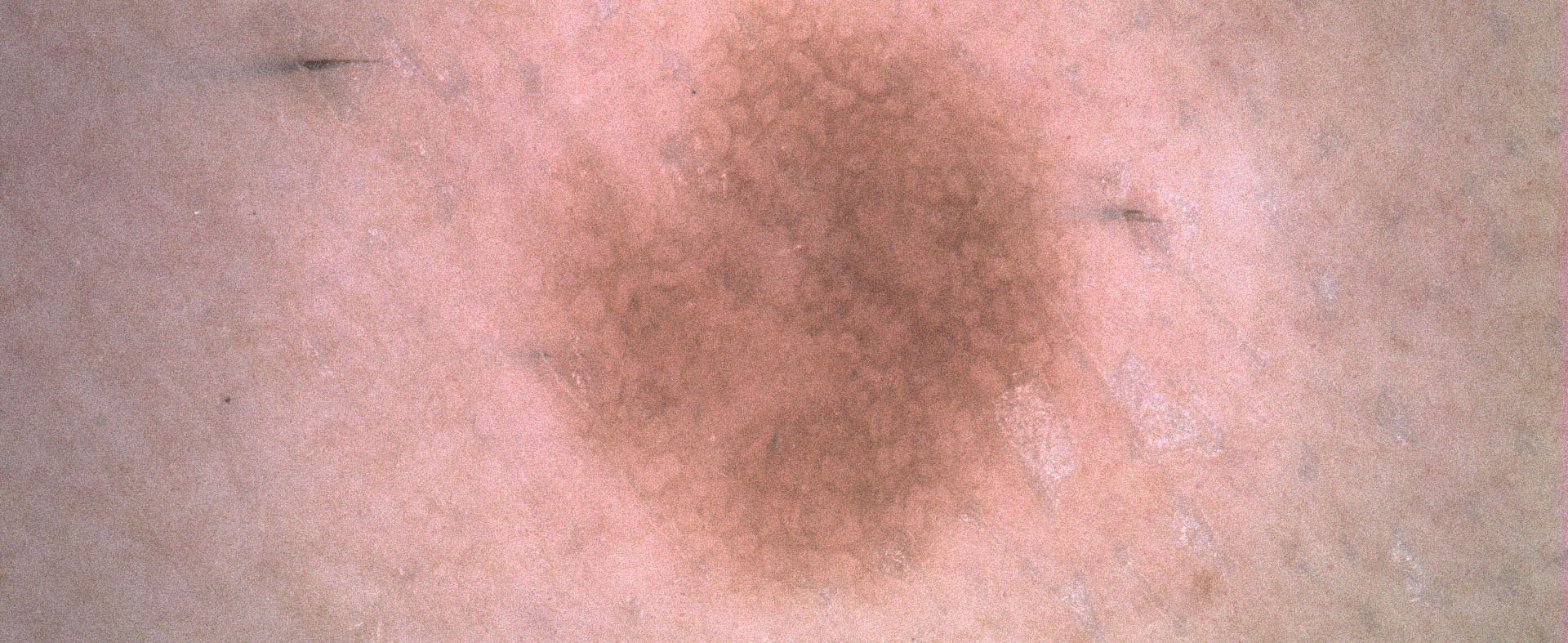
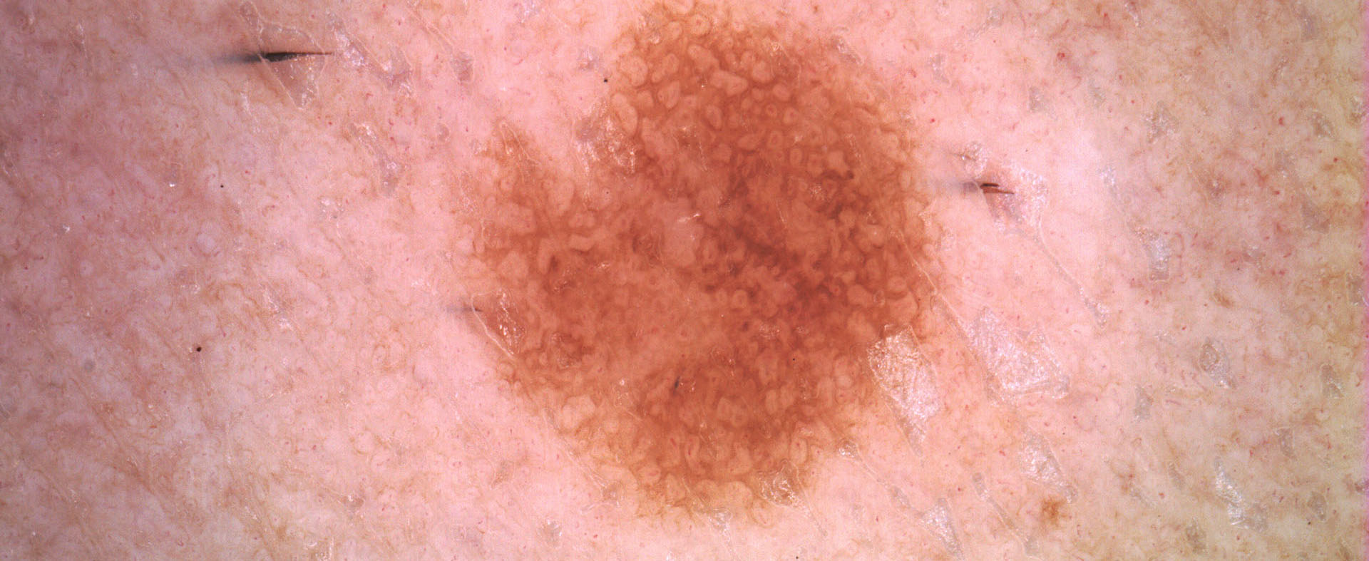
Color balance
Sometimes observe the dermoscopic images with a different light helps to find small details that can be vital in diagnostic processes.
The Color Balance function of IRSKIN professional software is a standard algorithmic tool that balances the color depth of the captured images.
Since editing the images, it can be used only in the visualization phase, simply by checking the Color Balance box.

Before using
color balance

After using
color balance

Before using
color balance

After using
color balance
Razor
Frequently dermoscopic images of skin lesions appear partially covered by hair, which makes the evaluation of the mole’s features more complicated for the clinician.
A possible solution is to cut or eradicate possible hair present on the skin lesion, but this is not always desireable.
The IRSkin software offers a different solution, the Razor function, which attempts an algorithmic elimination of the hair.

Before using razor

After using razor
Segmentation
IRSkin Segmentation determines the edge of the lesion, identified by a Jordan curve that separates the lesion’s area from the background.
The segmentation automatically enlights the boundary of the skin lesion in real time, detecting the boundary of the lesion, its center, its orientation and the ellipse that approximates the entire mole.


Analysis
The Segmentation function activates the ABCD Analysis of a skin lesion image.
IRSkin’s ABCDE analysis provides the physician with quantitative information on the morphological and pigmentary characteristics of the skin lesion.
The ABCD analysis functions, together with the possibility of analysing the Evolution of a lesion by means of the Body Map, provide quantitative and qualitative information that may help the doctor improving its standard ABCDE analysis.
ABCD Analysis View will appear on the screen, together with the following numerical parameters:

ASYMMETRY:
Major Axis Symmetry ( percentage of symmetry of the segmented skin lesion with respect to its major axis of inertia)
Minor Axis Symmetry (percentage of symmetry of the segmented skin lesion with respect to its minor axis of inertia)
Central Symmetry (percentage of symmetry of the segmented skin lesion with respect to its barycenter)
Avg Symmetry (average of the three previous Symmetries)BOUNDARY PARAMETERS:
Shape Factor (numerical quantity measuring the complexity of the boundary of a shape with respect to its area: it is proportional to the ratio between the Area of the skin lesion and the square of its Perimeter. Small values of the Shape Factor indicate that the lesion has an articulated boundary)
Haralick’s Circularity (statistical quantity measuring the resemblance of a shape with a circle. Large values of the Haralick’s circularity indicate the the lesion has a high circularity)
Ellipticity (numerical quantity measuring the resemblance of a shape with its inertial ellipse. Large values of Ellipticity indicate that the lesion is similar to its inertial ellipse)
Eccentricity (numerical quantity measuring the ratio between the inertial axes of a shape: large values of Eccentricity indicate an extended skin lesion)COLOR:
Color Prevalence (graphic indicating the distribution of the prevailing colors of the skin lesion)
Color Zones visualization (of the prevailing colors of the skin lesion and of its inertial axes)
Color Entropy (statistical quantity, defined by the IRSkin Team, measuring the color variations of the skin lesion among its four quarters)DIMENSIONAL PARAMETERS (expressed in millimeters):
Diameter (maximal distance between two points of a skin lesion)
Area (measure of the area covered by the skin lesion)
Perimeter (measure of the length of the boundary of the skin lesion)
Length (measure of the of length of the major axis of the skin lesion)
Height (measure of the length of the minor axis of the skin lesion)
DIGITAL TRICHOSCOPY
Using the Dermoscopic Camera on the scalp, various parameters such as hair structure, dimensions, circulation around the follicle or abnormal structures can be analyzed without any annoyance for the patient. By storing the images, the software allow to appreciate efficacy of the therapies and the evolution of disorders.

VIDEO CAPILLAROSCOPY
Capillary morphology and blood flow can be analyzed in a simple, and easily repeatable way. Placing the Dermoscopic Camera on the area allows to acquire and store images, the follow up of the damage of the microcirculation and appreciate the efficacy of the therapy.
